Chapter outline
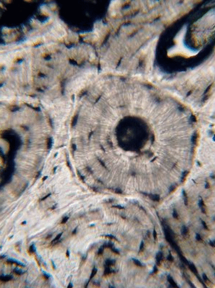
Bone tissue is deposited in layers by osteoblasts. These cells become trapped within lacunae between layers of bone tissue, and differentiate into osteocytes. Bone tissue can either be compact and made of osteons, or spongy bone made of trabeculae. Bone tissue is almost entirely ECM, composed of:
| 66% | Mineral: Calcium Hydroxyapatite |
| 33% | Protein: Collagen |
| 96% | Mineral: Calcium Hydroxyapatite |
| 4% | Protein: Amelogenins and enamelins, not collagen |
| 70% | Mineral: Calcium Hydroxyapatite |
| 30% | Protein: Collagen, mostly |
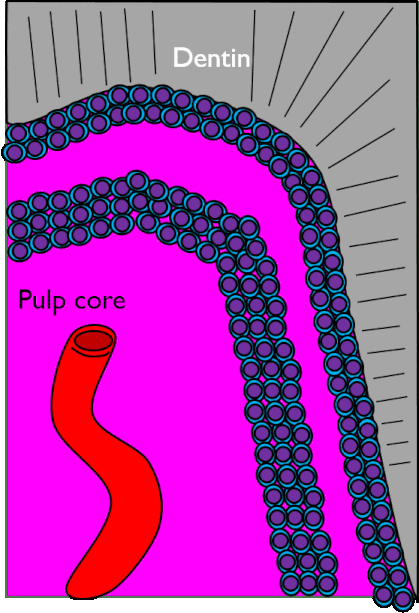
| Odontoblast layer | Odontoblast cell bodies |
| Cell-free zone | Not actually free of cells, they just aren't visible |
| Cell-rich zone | Cells are visible and densely packed |
| Pulp core | Location of capillaries, nerve endings and lymphatic vessels |
| 50% | Mineral: Calcium Hydroxyapatite |
| 50% | Protein: Collagen and proteoglycan mixture |
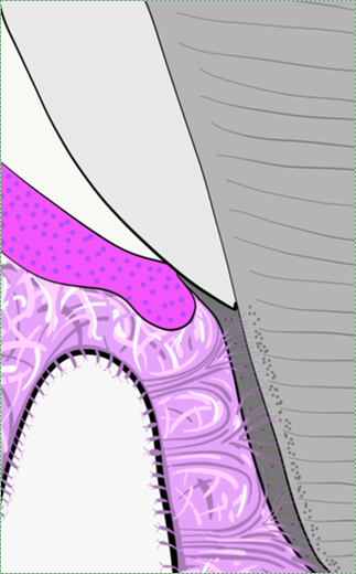
| Enamel | 96% | Mineral: Calcium Hydroxyapatite |
| 4% | Protein: Amelogenins and enamelins, not collagen | |
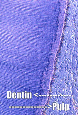
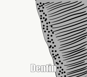
| Dentin | 70% | Mineral: Calcium Hydroxyapatite |
| 30% | Protein: Collagen, mostly | |
| Cementum | 50% | Mineral: Calcium Hydroxyapatite |
| 50% | Protein: Collagen and proteoglycan mixture | |
| Bone | 66% | Mineral: Calcium Hydroxyapatite |
| 33% | Protein: Collagen |